Category

Surgeries

5 Minutes Prostate Surgery With Our Rezum Expert Dr. Fouad Khoury


52 years old male patient:

45 gr prostate with failure to improve symptoms on medical treatment.

Taking into consideration the patient’s age and sexual activities; he was insisting on undergoing a procedure to relieve his symptoms while preserving EJACULATION.

A 5 minutes prostate procedure (REZUM) as shown in the video,  was done by our Rezum Expert Dr.Fouad Khoury under local anesthesia.

There is no need for the patient to sleep in the hospital, or to be hospitalized.

Prostate treatment in Middle East 

Rezum water vapor

Recently approved by the FDA in 2015, makes it possible to perform a prostate surgery in 5 minutes while preserving EJACULATION AND SEXUAL FUNCTION.

This operation consists of throwing steam through an instrument into the enlarged area of ​​the prostate; the vapor carries energy that will be diffused into the molecules which will be killed and discarded by the immune system.

It lasts 5 to 10 minutes under local anesthesia and the number of vapor ejections depends on the volume of the prostate.

Benefits of REZUM

– The procedure doesn’t cause complications, no need to be hospitalized.

– Return to daily activities in a few days.

– Relatively painless.

– Improvement of symptoms after 2 weeks.

– Symptoms continue to improve for up to 3 months.

– A good prognosis of up to 15 years.

– This procedure doesn’t cause sexual dysfunctions.

After this operation, the doctor can give an antibiotic for a few days to prevent infection.


Facts about bladder prolapse in Middle East


Bladder Prolapse

What’s bladder prolapse?

A cystocele or a prolapsed bladder occurs when ligaments that hold your bladder up and the muscle between a woman’s vagina and bladder are weak; for this reason, the bladder will fall into the vagina.

There are three grades of cystocele:

– Grade 1: The bladder falls only a short way into the vagina.

– Grade 2: The bladder falls to the opening of the vagina.

– Grade 3: The bladder comes out of the opening of the vagina; it’s a severe grade.

Causes and Risk factors

There are several causes and risk factors:

– Pregnancy and childbirth: Women who have had a vaginal delivery, multiple pregnancies, or whose infants had a high birth weight have a higher risk of anterior prolapse; because the pregnancy and the delivery may involve straining the muscles of the floor of the pelvis.

– Age:  especially after menopause, when the production of estrogen decreases; so the muscles around the vagina are not strong anymore.

– Hysterectomy.

– Family history or genetic factor (some women are born with weak connective tissues).

– Obesity: Women who are obese are at higher risk of anterior prolapse.

– Constipation.

– Chronic coughing.

– Lifting heavy objects.

– Smoking.

What are the symptoms of a cystocele?

– Seeing something bloating through the vaginal opening.

– Difficulty to urinate.

– Feeling the need to urinate frequently.

– Frequent urinary tract infections.

– Pain in the pelvic area or lower back.

– Painful sex.

– Difficulty to insert tampons or applicators.

How is bladder prolapse diagnosed?

Prolapsed Bladder can be diagnosed with a clinical history and a pelvic exam. The exam may be done while you are lying down, straining or pushing, or standing; the doctor may measure the severity of the prolapsed bladder by seeing in which part of the vagina the bladder has fallen.

Other tests and imaging studies may also be done to check the pelvic floor, and confirm the diagnosis of a prolapsed bladder:

– Cystoscopy: a long tube is passed through the urethra to examine the bladder.

– Urodynamics: to measure the capacity of the bladder to hold and release urine.

– X-rays

– Ultrasound

– MRI

How to treat the prolapsed bladder?

1- No treatment if the prolapse is:

– Not causing you problems.

– Not blocking your urine flow.

 2- Behavior therapy

– Kegel exercises (which help the muscles of the pelvic floor to be strong).

– Pelvic floor physical therapy.

– Pessary: a vaginal support device to hold the bladder in place.

3- Estrogen replacement therapy

4- Weight loss

5- Surgery:

A moderate or severe prolapsed bladder may require reconstructive surgery to move the bladder into a normal position; and it can be performed through the vagina or the abdomen, under a spinal or general anesthetic.

The surgical treatment is the best therapy for bladder prolapse; but if a woman is planning for a pregnancy, surgery is contraindicated.

The patient usually goes home the day of the surgery, and the recovery time typically takes four to six weeks.

Follow the link for more information about bladder prolapse treatment.

How can a cystocele be prevented?

– Maintain a healthy weight.

– Avoid lifting heavy objects.

– Treat constipation.

– Stop smoking.


Diagnosis and Treatment for Bladder cancer

How to diagnose Bladder cancer?

There are many tests that help to diagnose bladder cancer.

– Cystoscopy:
This procedure is required to identify and diagnose bladder cancer. A cystoscope is inserted under local or general anesthesia into your bladder from the urethra to view the inside of the bladder and take a Biopsy that will be examined in the laboratory.

– Urine cytology:
A sample of your urine is analyzed under a microscope to check some tumor markers in the urine.

After confirming that you have bladder cancer, your doctor may recommend some imaging tests to determine whether your cancer has spread to your lymph nodes or to other areas of your body.

– CT scan of the abdomen and pelvis to determine if there is any propagation of the tumor outside of the bladder.

– Magnetic resonance imaging (MRI) scan:
MRI scans show detailed images of soft tissues in the body, like CT scans; but by using radio waves and strong magnets instead of x-rays.

– PET scan: can detect cellular changes in organs and tissues earlier than CT and MRI scans; by injected a radioactive chemical.

– Chest X-ray is performed to detect if any cancer has propagated to the lungs.

– Bone scan may be performed to look for metastasis to cancer in the bones.

Follow the link for more information about the diagnosis of bladder cancer.

How to treat Bladder cancer?

• Surgery
• Chemotherapy
• Radiation therapy
• immunotherapy for superficial cancers
Sometimes, combinations of these treatments will be used.

-Surgery:
Transurethral resection of bladder tumor (TURBT):
TURBT is a procedure to diagnose bladder cancer and to remove cancers in the cells of the bladder lining (no muscle-invasive cancers). It is performed during a cystoscopy by using an electric current to cut away or burn away cancer.

Cystectomy:
– A radical cystectomy is an operation to remove all the bladder and the surrounding lymph nodes when the tumor has spread beyond the bladder. In men, radical cystectomy consists of removing the prostate and seminal vesicles; and in women, a radical cystectomy may involve the removal of the uterus, ovaries, and part of the vagina. It can be done using Robotic Surgery.
– Partial cystectomy is the removal of a section of the bladder when the tumor is only in one region of the bladder. Chemotherapy or radiation therapy is often used in combination with this procedure.

Chemotherapy :
By using a drug to kill or stop the reproduction of cancerous cells.
Chemotherapy drugs can be delivered
– Intravenously: Intravenous chemotherapy is frequently used before cystectomy to have a high chance of curing cancer, or can be used to kill cancer cells that might remain after surgery.
– Intravesically directly into the bladder.
It depends on the stage of cancer.

Radiation therapy:
Destroys the DNA of cancer cells by using powerful energy, like X-rays and protons.
It can be used in fusion with surgery or chemotherapy. Radiation therapy can be delivered externally or internally.

Immunotherapy:
Immunotherapy is a drug treatment that helps your immune system to fight cancer, by using the body’s own immune system.
Immunotherapy can be performed:
• Directly into the bladder: Might be required after TURBT for small bladder cancers that haven’t grown into the deeper muscle layers of the bladder. This treatment uses bacillus Calmette-Guerin (BCG), which causes an immune system reaction; so the body can kill the tumor bladder cells.
• Intravenously: Immunotherapy can be performed intravenously for bladder cancer that’s advanced or that comes back after initial treatment.

Follow the link to know about BCG Therapy.


Maintenance Therapy For Bladder Cancer

Maintenance Therapy For Bladder Cancer … What is the role of BCG?

Bladder cancer can come back; for this reason, the patient needs follow-up tests for years after finishing the treatment.

The best follow-up treatment is the BCG: Bacillus Calmette-Guérinthat used since 1920 to attenuated the action of the tubercle bacilli that causes tuberculosis.

The action of BCG

The local immune response is closely linked to the interaction of three systems: the patient, the BCG (Mycobacteria), and the tumor. This interaction will give rise to a cascade of immunological events, some of which will be essential for the protective action of BCG against tumor recurrence and progression. There are three phases in the immune response to BCG. First, the BCG adheres to the urothelium and then is phagocytosed by antigen-presenting cells; this phase corresponds to the early release of inflammatory cytokines. These cytokines could be involved in certain undesirable effects but they could also participate in cytotoxic phenomena. The second phase is the recognition of bacterial antigens by CD4 helper lymphocytes. This cellular activation will lead to the third phase which is the amplification of cytotoxic populations capable of killing tumor cells. All of these cells also produce cytokines that help regulate the immune response.

Indications of BCG:

BCG (powder and solvent for suspension for intravesical use) is used to :

-curative treatment of urothelial carcinoma in situ.

-prophylactic treatment of relapses of urothelial carcinoma limited to the mucosa, no muscle-invasive urothelial carcinoma, urothelial carcinoma in situ.

Protocol of injection of BCG:

2-3 weeks after TURBT ( weekly for 6 weeks).

After a cystoscopy: Maintenance therapy consists of 3 treatments at weekly intervals given for a minimum of 1 year
up to 3 years; every 3 months for 2 years, then every 6 months for 2 years, and finally yearly.

What are the side effects of BCG?

It is common for patients to experience flu-like symptoms for 2–3 days after the treatment.

Other side effects include:

-Trouble to empty your bladder.

-Blood in your urine, dark urine.

-Urinary tract infections.

-Pain when you urinate.

-Vomiting, pain in the upper part of the stomach.

-Trouble breathing.

-Fatigue.

-Signs of a penis infection: burning, itching, odor, discharge, pain, tenderness, redness or swelling of the genital or rectal area, fever, not feeling well.

-Yellowing of your skin or eyes.

After the treatment (after 4 to 6 hours), the patient may present bladder symptoms: sudden need to urinate, frequent urination, stomach discomfort, bloating, and possibly loss of bladder control. If these symptoms last for more than 2 days; you should consult your urologist.

Follow the link for more information about BCG.


Facts About Bladder Cancer in Middle East

Bladder Cancer

Definition of the Bladder :

The bladder is an empty muscular organ that stores urine until it is discharged out of the body through the urethra, and which is located in your lower abdomen.

Where is bladder cancer located?

Bladder cancer begins in the cells of the bladder lining, most commonly in the urothelial cells; this cancer grows and forms a tumor.
Urothelial cells are found in your kidneys and the ureters which is the connection between your kidneys and your bladder.
Urothelial cancer can occur in the kidneys and ureters, too, but it’s more common in the bladder.
When a mutation occurs in the DNA of the bladder cells; cancer begins.

Types of bladder cancer

Different types of cells in your bladder can become cancerous; the type of bladder cancer depends on where the tumor’s cells begin. Doctors use this information to determine which treatment is the best for you.
The 3 main types of bladder cancer are:

– Urothelial carcinoma

Urothelial carcinoma or transitional cell carcinoma begins in the urothelial that line the inside of the bladder. Urothelial cells dilate when your bladder is full and contract when your bladder is empty. These same cells existent inside of the ureters and the urethra; so cancer can form in those places. Urothelial carcinoma is the most frequent type of bladder cancer in the United States.

– Squamous cell carcinoma
This type of cancer is triggered by chronic irritation of the bladder due to repeated urinary tract infections, especially in countries where the parasitic infection is the cause of bladder infections; and due to long-term use of a urinary catheter.

– Adenocarcinoma
Adenocarcinoma begins in cells that elaborate mucus-secreting glands in the bladder, and it’s very rare.
Some bladder cancers involve more than one type of cell.

Stages of Bladder Cancer…TNM staging system

This system is used by doctors to determine the stage of bladder cancer (Tumor, Nodule, and Metastasis).

Bladder cancer can be limited to the lining of the bladder or invasive (penetrating the bladder wall and possibly spreading to nearby organs or lymph nodes).

– Invasive bladder tumors can be classified from T2 (spread to the main muscle wall below the mucosa of the bladder) to T4 (tumor is extended beyond the bladder to nearby organs or the pelvic sidewall).

– Lymph node involvement classifies from N0 (no cancer in lymph nodes) to N3 (cancer in many lymph nodes, or in one or more bulky lymph nodes larger than 5 cm).

– M0 means the absence of metastasis outside of the pelvis, M1 means that the tumor has metastasized outside of the pelvis.

Follow the link for more information about stages of Bladder Cancer.

Signs of bladder cancer

Bladder cancer has no specific symptoms, so when the patient has: blood in the urine, pain during urination, frequent urination, or difficulty urinating; he should visit his urologist.

Risk factors

• Smoking
• Increasing age: especially when the patient is older than 55.
• Men have a high risk than women to develop bladder cancer.
• Certain chemicals products.
• Previous cancer treatment; a patient treated with the anti-cancer drug cyclophosphamide is at high risk of developing bladder cancer. People who already received radiation treatments focused on the pelvis for previous cancer have a higher risk of developing bladder cancer.
• Chronic bladder inflammation: due to repeated urinary infections, or long-term use of a urinary catheter; may expose the patient to bladder cancer.
• Personal or family history of bladder cancer.


Follow the link for more information about the diagnosis and treatment of bladder cancer.

Penile Prosthesis the BEST surgical treatment for ED in Middle East

Penile prosthesis

Definition

A penile prosthesis or Penile Implant is the best surgery that helps men with erectile dysfunctions when other treatments failed.

This procedure involves placing a prosthetic device or penile implant inside the penis and scrotum; so the patient can get a sufficient erection for sexual activities. And it lasts for 45 minutes to 1 hour under general or spinal anesthesia.

Penile implants are required when the medications are not efficient, and in some severe cases like Peyronie’s disease (fibrous scar tissue that develops on the penis and causes curved painful erections).

Follow the link for more information about Erectile Dysfunction.


Types of penile implants:

The patient should speak with his urologist to determine which implant is the best for him.


Type of penile implant

Advantages

Disadvantages

Three-piece inflatable

 -Natural and rigid erection.

-Provides flaccidity when deflated.


 -The implant may sometimes not be effective (because of the large number of its parts).

 -Requires the presence of a reservoir in the abdomen.


Two-piece inflatable

 -Provides flaccidity when deflated.

 -The fluid reservoir is part of the pump.

 -The erection is not firm enough.

Semirigid rod

 -Low chance of malfunction. ( due   to   the absence of the reservoir   and the   pump)

 – Easy to use.

 -Can be difficult to conceal under clothing.

 -a penis that is always slightly rigid.

 -possible difficulty with urination.


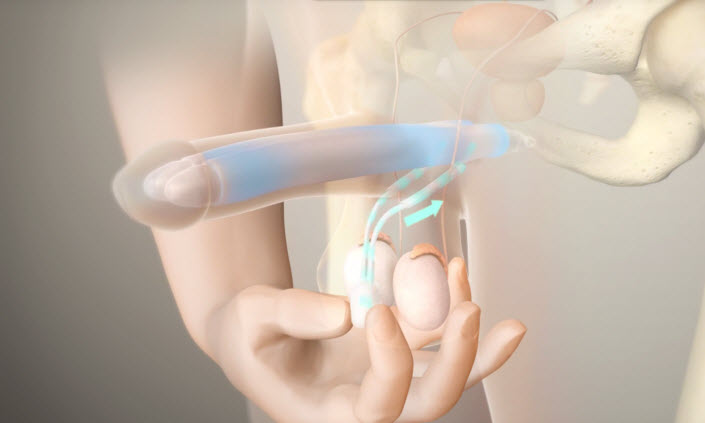
The inflatable implants required a pump inside the scrotum; so the patient should squeeze the pump to achieve an erection; the pump is located under the loose skin of the scrotal sac, between the testicles.

The device contains two chambers, and when the chambers are inflated by the pump; the patient then has an erection; when the patient is finished, he can deflate the device.

Follow the link for more details about the types of penile implants.


Which type of implant does the doctor choose?

There are several factors that the doctor put into consideration before deciding which implant will be the most suitable, including:

-The age of the man.

-Size of the penis, glans, and scrotum.

-Any history of previous abdominal or pelvic surgery.

-The presence of colostomy.

-A history of a kidney transplant.

-Whether or not the penis is circumcised.

-Health and well-being.

 Penile implants don’t increase sexual desire or sensation.


Who should not get an implant?

-patient with uncontrolled diabetes.

-presence of a pulmonary or urinary infection.

-bladder obstruction

-when the erectile dysfunction is the result of a relationship conflict (the cause should be medical).


Penile implant = LOW RISK

– Low risk of infection (1-3%).

-low risk of mechanical failure (95% working at 5 years).


Some TIPS before and after the surgery

Before the surgery you should:

– stop taking aspirin and anti-inflammatory drugs before 7 to 10 days.

– Stop eating or drinking after midnight before your surgery.

– Shave the surgery site.

After the surgery:

– Physical and sexual activities can be resumed after 4 or 6 weeks.

-the patient should take an antibiotic to prevent infection; and medications to ease the pain.

You should call your doctor if:

fresh and ongoing bleeding, significant discoloration of the penis, high fever, unable to urinate, cannot control pain, spreading redness, continuous drainage from the wound, progressive swelling of the penis, scrotum, or incision site.


Is the Prosthesis noticeable?

Men who have undergone the prosthesis surgery can notice the small surgical scar where the bottom of the penis meets the scrotal sac, or in the lower abdomen just above the penis, other people probably will be unable to know that a penile implant exists.


Finally, we believe that the penile implant is effective in its ability to restore the patient’s capacity to engage in sexual activities and regain what was lost due to a medical issue.

And the patient should not forget that ejaculation is not affected by this procedure.

Laparoscopic Radical Nephrectomy-14 cm tumor

This movie features a challenging laparoscopic left radical nephrectromy for a huge tumor compressing the aorta and overlying the kidney pedicle.

The procedure was successfully performed by Dr. Fouad Khoury with minimal blood loss and optimal post-operative results.


First Complex Robotic Assisted Partial Nephrectomy

Robotic Assisted Partial Nephrectomy


What is Robotic Assisted Partial Nephrectomy?

Robotic partial nephrectomy surgery consists of removing a part of a kidney, to treat cancer by preserving as much healthy kidney tissue as possible.

Who needs a robotic partial nephrectomy?

-Patient with small kidney tumor (<4 cm in size).

-Patient with risk of kidney failure and the need of dialysis when the procedure consists of removing all the kidney.

Kidney Tumors between 4 and 7 centimeters can be treated with robotic partial nephrectomy if they are located in certain areas.

Some Tips before the surgery:

-Stop taking blood thinners 5 to 7 days before the surgery.

-Anti-inflammatory medications and certain vitamin supplements can cause increased bleeding, so the surgeon and the anesthetist should be informed about all your medications.

-It’s very necessary to stop smoking few days before the procedure.

-Not eat or drink anything after midnight the night before your surgery, to prevent anesthesia complications.

During a robotic partial nephrectomy:

This procedure is performed under general anesthesia, by making small cuts in the abdomen; so the robotic surgical equipment and camera can be inserted.

Carbon dioxide gas is used to inflate the abdominal cavity to leave space for the manipulation of the surgical equipment and camera to access the cancerous tissues.

While the surgeon is performing the procedure by manipulating the robot, the blood flow to the cancerous kidney is totally stopped; so the kidney can be dissected and the cancerous portion is detached from the tissue. The tumor is totally removed from the body and the surgeon sews the remaining section of the healthy kidney.

After a robotic partial nephrectomy:

– Take analgesics to reduce post-operative pain.

-Walk after a few days of surgery to promote good blood circulation and prevent pneumonia.

-Start with a liquid diet for a few days after the operation.

Robotic partial nephrectomy VS Laparoscopic partial nephrectomy

Robotic partial nephrectomy is more efficient than laparoscopic partial nephrectomy because the da Vinci surgical system has a lot of advantages.

Follow the link for more information about the difference between robotic partial nephrectomy and laparoscopic partial nephrectomy.


Robotic-assisted right partial nephrectomy for a complex 8 cm tumor impinging on the collecting system in an elderly patient with chronic renal insufficiency (eGFR of 40 mL/min)

Laparoscopic Ureterolithotomy – 2.5 cm Proximal Ureteral Stone

Laparoscopic ureterolithotomy for a large 2.5cm proximal ureteral stone. After mobilization of the colon, identification of the ureter and dissection of the inflammatory region when the stone is impacted. Opening of ureter using cold cut in order to obtain a neat incision and prevent thermal damage to the ureter.

Extraordinary Laparoscopic Pyeloplasty (Left)

Dr Fouad Khoury, a referral urologist in Lebanon and the Middle East, performing pyeloplasty for UPJ stenosis. This video features step by step approach along with captions for successful and smooth completion of pyeloplasty operation, with antegrade intra operative JJ insertion.

Read More